Medical Ultrasound Imaging
Friday, 4 April 2025
'Probe' p19 Searchterm 'Probe' found in 121 articles 11 terms [ • ] - 110 definitions [• ] Result Pages : •  From Philips Medical Systems;
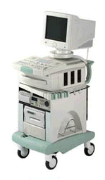
Introduced in June 2005, 'one of the less expensive and more dedicated' ultrasound systems.
Device Information and Specification
CONFIGURATION
LCD monitor
Broadband, convex, linear,
digital beamformer and focal tuning IMAGING OPTIONS
OPTIONAL PACKAGE
DICOM, etc.
STORAGE, CONNECTIVITY, OS
HDD, CD, USB, optionalMOD and DICOM 3.0
DATA PROCESSING
256-digitally processed channels
H*W*D inch.
58 * 20 * 32
WEIGHT
135 lbs.
•
(HIFU / FUS) High intensity focused ultrasound is used in thermotherapy or thermoablation e.g., for the treatment of benign prostate hyperplasia or under study for the treatment of cancer. An applied ultrasound probe (see transrectal sonography) focuses sound waves at one spot, elevating the tissue temperature to a point that the tissue destroys. Generally, lower frequencies (from 250 kHz to 2000 kHz) are used than for medical diagnostic ultrasound, but significantly higher time-averaged intensities. See also Magnetic Resonance Guided Focused Ultrasound, Low Intensity Pulsed Ultrasound, and Lithotripsy. Further Reading: Basics: •
Linear array transducer elements are rectangular and arranged in a line. Linear array probes are described by the radius of width in mm. A linear array transducer can have up to 512 elements spaced over 75-120 mm. The beam produced by such a narrow element will diverge rapidly after the wave travels only a few millimeters. The smaller the face of the transducer, the more divergent is the beam. This would result in poor lateral resolution due to beam divergence and low sensitivity due to the small element size. In order to overcome this, adjacent elements are pulsed simultaneously (typically 8 to 16; or more in wide-aperture designs). In a subgroup of x elements, the inner elements pulse delayed with respect to the outer elements. The interference of the x small divergent wavelets produces a focused beam. The delay time determines the depth of focus for the transmitted beam and can be changed during scanning. Linear arrays are usually cheaper than sector scanners but have greater skin contact and therefore make it difficult to reach organs between ribs such as the heart. One-dimensional linear array transducers may have dynamic, electronic focusing providing a narrow ultrasound beam in the image plane. In the z-plane (elevation plane - perpendicular to the image plane) focusing may be provided by an acoustic lens with a fixed focal zone. Rectangular or matrix transducers with unequal rows of transducer elements are two-dimensional (2D), but they are termed 1.5D, because the number of rows is much less than the number of columns. These transducers provide dynamic, electronic focusing even in the z-plane. See also Rectangular Array Transducer. •  From ESAOTE S.p.A.;
'Megas GPX means all digital, all configurations, all probes feature a high standard level of quality and extended versatility. Based on digital beamformer technology, Megas GPX's functions can be easily updated to explore the new frontiers of the ultrasound world and give you the most flexible and powerful tool to make even the most difficult diagnoses.' •
Standard scanners allow visualizing microbubbles on conventional gray scale imaging in large vascular spaces. In the periphery, more sensitive techniques such as Doppler or non-linear gray scale modes must be used because of the dilution of the microbubbles in the blood pool. Harmonic power Doppler (HPD) is one of the most sensitive techniques for detecting ultrasound contrast agents. Commonly microbubbles are encapsulated or otherwise stabilized to prolong their lifetime after injection. These bubbles can be altered by exposure to ultrasound pulses. Depending on the contrast agent and the insonating pulse, the changes include deformation or breakage of the encapsulating or stabilizing material, generation of free gas bubbles, reshaping or resizing of gas volumes. High acoustic pressure amplitudes and long pulses increase the changes. However, safety considerations limit the pressure amplitude and long pulses decrease spatial resolution. In addition, lowering the pulse frequency increases destruction of contrast bubbles. However, at low insonation power levels, contrast agent particles resist insonation without detectable changes. Newer agents are more reflective and will usually allow gray scale imaging to be used with the advantages of better spatial resolution, fewer artifacts and faster frame rates. Feasible imaging methods with advantages in specific acoustic microbubble properties: Resonating microbubbles emit harmonic signals at double their resonance frequency. If a scanner is modified to select only these harmonic signals, this non-linear mode produces a clear image or trace. The effect depends on the fact that it is easier to expand a bubble than to compress it so that it responds asymmetrically to a symmetrical ultrasound wave. A special array design allows to perform third or fourth harmonic imaging. This probe type is called a dual frequency phased array transducer. See also Bubble Specific Imaging. Result Pages : |
Medical-Ultrasound-Imaging.com
former US-TIP.com
Member of SoftWays' Medical Imaging Group - MR-TIP • Radiology TIP • Medical-Ultrasound-Imaging
Copyright © 2008 - 2025 SoftWays. All rights reserved.
Terms of Use | Privacy Policy | Advertise With Us
former US-TIP.com
Member of SoftWays' Medical Imaging Group - MR-TIP • Radiology TIP • Medical-Ultrasound-Imaging
Copyright © 2008 - 2025 SoftWays. All rights reserved.
Terms of Use | Privacy Policy | Advertise With Us
[last update: 2023-11-06 01:42:00]


