Medical Ultrasound Imaging
Sunday, 20 September 2026

 Info Sheets  Out- side     | 'Probe' p16 Searchterm 'Probe' found in 121 articles 11 terms [ • ] - 110 definitions [• ] Result Pages : •
Cardiac ultrasound, also known as echocardiography or echocardiogram, is used to provide several different levels and types of heart testing. Cardiac ultrasound utilizes the same ultrasound principles as used for obstetric and gynecologic evaluations of pregnant women, gallbladder ultrasound and other abdominal structures. The ultrasound is directed out of a hand held probe which can be moved to image the heart from different positions. Additionally, so that heart events can be timed, ECG leads are placed on the chest. The reflected wave is converted into an actual image of the heart and displayed in a real-time mode or M-mode ultrasound format. M-mode recordings permit measurement of cardiac dimensions and detailed analysis of complex motion patterns depending on transducer angulations. Also the time relationships with other physiological variables such as ECG, heart sounds, and pulse tracings, can be recorded simultaneously. A stress echocardiogram provides information about the cardiac performance. Two-dimensional tomographic images of selected cardiac sections give more information than M-mode about the shape of the heart and also show the spatial relationships of its structures during the cardiac cycle (diastole to systole). See also M-Mode Echocardiography, and Myocardial Contrast Echocardiography.  Further Reading: News & More:
• 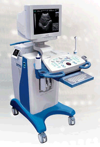 From CHISON MEDICAL IMAGING CO., LTD.;
PC-based scanner for abdominal, Obs/Gyn, small parts, urology, cardiology, proctology, pediatrics with four 4-step multi-frequency probes. •
The cable, which connects the probe (also called handle) with the ultrasound machine, contains from 48 to 256 micro-coaxial cables, is about 2 m long and is one of the most expensive parts of the system. •
(CPA) Color power angio is a Doppler measurement, which employs the Doppler effect to assess whether blood is moving towards or away from the probe. By calculating the frequency shift of a particular sample volume, its speed and direction can be determined and visualized. This is particularly useful in cardiovascular studies and essential in many areas such as determining reverse blood flow in the liver vasculature in portal hypertension. The CPA information is displayed as a color image superimposed on the 2D gray scale image. See also Color Power Doppler. •
Convex transducers are today standard on every new scanner. A convex surface allows the scanning of a larger area with a smaller array. The method of focusing and beam sweeping of a convex or curvilinear / curved array is similar to a linear array transducer, except of the shape of the probe and the sector format of the created image. The better fit to the body, caused by the curved shape with smaller convex contact surface, and the wider field of view further from the transducer face are advantages in abdominal ultrasound. However, also a convex array is often too large to image the heart when probing between the ribs. Caused by combining a large field of view with smallest array size, phased array transducers are the best choice in cardiac ultrasound. See also Curved Transducer. Result Pages : | Share This Page     Look Ups |
Medical-Ultrasound-Imaging.com
former US-TIP.com
Member of SoftWays' Medical Imaging Group - MR-TIP • Radiology TIP • Medical-Ultrasound-Imaging
Copyright © 2008 - 2025 SoftWays. All rights reserved.
Terms of Use | Privacy Policy | Advertise With Us
former US-TIP.com
Member of SoftWays' Medical Imaging Group - MR-TIP • Radiology TIP • Medical-Ultrasound-Imaging
Copyright © 2008 - 2025 SoftWays. All rights reserved.
Terms of Use | Privacy Policy | Advertise With Us
[last update: 2025-04-05 02:38:00]