Medical Ultrasound Imaging
Wednesday, 2 April 2025
'Display' p14 Searchterm 'Display' found in 81 articles 1 term [ • ] - 80 definitions [• ] Result Pages : •
Diagnostic ultrasound imaging has no known risks or long-term side effects. Discomfort to the patient is very rare if the sonogram is accurately performed by using appropriate frequencies and intensity ranges. However, the application of the ALARA principle is always recommended.
There are reports of low birth weight of babies after applying more than the recommended ultrasound examinations during pregnancy. Women who think they might be pregnant should raise this issue with the doctor before undergoing an abdominal ultrasound, to avoid any harm to the fetus in the early stages of development. Since ultrasound is energy, sensitive tissues like the reproductive organs could possibly sustain damage if vibrated to a high degree by too intense ultrasound waves. In diagnostic ultrasonic procedures, such damage would only result from improper use of the equipment. Possible ultrasound bioeffects:
•
•
Due to increasing of temperature, dissolved gases from microbubbles come out of the contrast solution.
The thermal effect is controlled by the displayed thermal index and the mechanical index indicates the risk of cavitation. An ultrasound gel is applied to obtain better contact between the transducer and the skin. This has the consistency of thick mineral oil and is not associated with skin irritation or allergy. Specific conditions for which ultrasound may be selected as a treatment may be attached with higher risks. See also Ultrasound Imaging Procedures, Fetal Ultrasound and Obstetric and Gynecologic Ultrasound. • View NEWS results for 'Side Effect' (7). •  From Kontron Medical SAS;
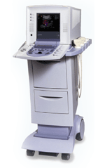
'The Sigma 330 is a versatile, digital, mobile Ultrasound System, upgradeable from 2D to Doppler, Color Flow Mapping and 3D. The Sigma 330 displays excellent image quality and superb Doppler and CFM sensitivity, employing ATEC™ (Advanced Tissue Echo Cancellation), a revolutionary technology developed by the Kontron Medical R&D Center, which removes tissue artifacts ('ghosting').' Specifications for this system will be available soon. •
The skinline is the ultrasonic penetration depth corresponding to the skin / transducer interface on the display.
•
Sonazoid™ is an ultrasound contrast agent (UCA) consisting of stabilized gas microbubbles in an aqueous suspension. Sonazoid™ has overcome the stability problems of first generation USCA and can produce myocardial perfusion images. Myocardial imaging using ultrasound contrast agents provides diagnosis of chronic heart disease and assessment of the coronary arteries and of the coronary blood flow reserve.
Sonazoid™ is taken up by healthy Kupffer cells in the liver and spleen, but break down in high amplitude ultrasound imaging modes such as color Doppler imaging. The bubble rupture produces a transient pressure wave, which results in a characteristic mosaic color pattern from tissues containing the microbubbles (induced acoustic emission). Liver tumors without Kupffer cells will not display the mosaic pattern and can therefore be identified easily.
Drug Information and Specification
RESEARCH NAME
NC100100
DEVELOPER
INDICATION -
DEVELOPMENT STAGE Development in USA and EU suspended
APPLICATION
-
TYPE
Microbubble
Lipid Stabilized (not disclosed)
CHARGE
Negative
Perfluorobutane
MICROBUBBLE SIZE
-
PRESENTATION
-
STORAGE
-
PREPARATION
Reconstitute with 2mL water
DO NOT RELY ON THE INFORMATION PROVIDED HERE, THEY ARE
NOT A SUBSTITUTE FOR THE ACCOMPANYING PACKAGE INSERT! •
The term 'sonogram' is often used interchangeably with 'ultrasound,' but it specifically refers to the resulting image or picture produced during a diagnostic ultrasound examination, also known as ultrasonography or sonography. It serves as a visual representation of the echoes detected by the transducer and provides detailed anatomical information about the area being examined. Sonograms are typically displayed on a monitor, printed on film, or stored digitally for further analysis and documentation by medical professionals such as sonographers and radiologists. They serve as invaluable diagnostic tools, aiding in the detection and evaluation of various medical conditions, as well as guiding interventions, ultrasound therapy, and treatment planning. The term 'ultrasound' itself refers to the technology used during a sonogram, but it also finds several other applications beyond medical imaging. These include echolocation, crack detection, and cleaning, among others. See also Ultrasound Imaging, Ultrasound Technology, Handheld Ultrasound, Ultrasound Accessories and Supplies, Environmental Protection and Ultrasound Elastography. Result Pages : |
Medical-Ultrasound-Imaging.com
former US-TIP.com
Member of SoftWays' Medical Imaging Group - MR-TIP • Radiology TIP • Medical-Ultrasound-Imaging
Copyright © 2008 - 2025 SoftWays. All rights reserved.
Terms of Use | Privacy Policy | Advertise With Us
former US-TIP.com
Member of SoftWays' Medical Imaging Group - MR-TIP • Radiology TIP • Medical-Ultrasound-Imaging
Copyright © 2008 - 2025 SoftWays. All rights reserved.
Terms of Use | Privacy Policy | Advertise With Us
[last update: 2023-11-06 01:42:00]


